Virochip Design: Part 9
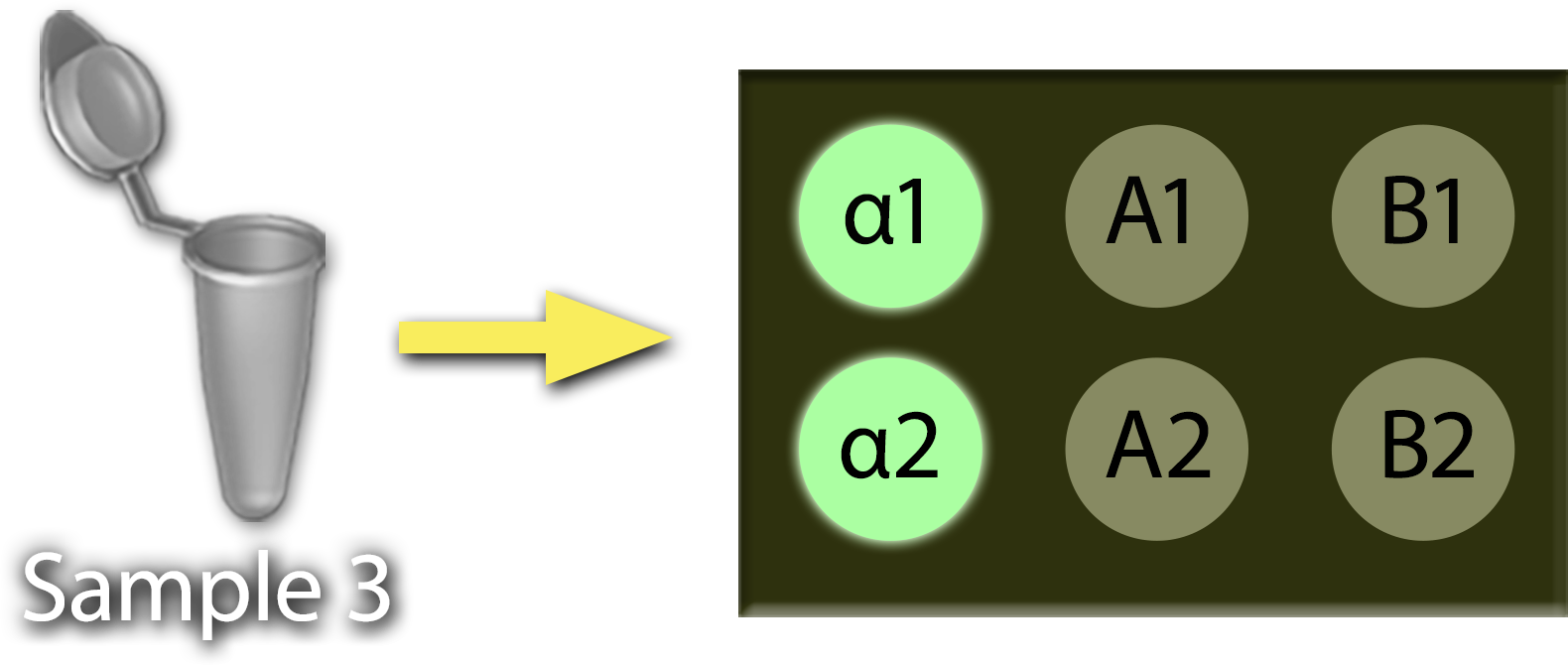
Running a different sample, you get a hybridization pattern like this. How would you interpret this result?


Running a different sample, you get a hybridization pattern like this. How would you interpret this result?

