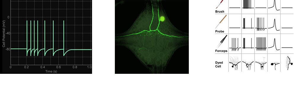
Overview of the Experiment

Through the following steps, you will explore how the leech's nervous system responds to different kinds of touch stimuli:

  1. Insert an electrode into a single neuron. Monitor the neuron's electrical response as you stimulate the skin with different tools.
  2. Inject the neuron with a fluorescent dye and observe its anatomy.
  3. Compare the electrical and anatomical data with published results to identify the neuron's type.
  4. Repeat the previous steps until you identify all the neurons.
A flowchart showing three images connected by arrows in a clockwise loop. The left image of action potentials is connected to the middle image showing a neuron filled with a fluorescent dye, which is connected to the right image showing an excerpt of the reference atlas of leech neuron activity and anatomy. The right image is also connected to the left image.
The left image is a graph labeled 'Time (s)' on the x-axis and 'Cell Potential (mV)' on the y-axis. The graph shows a single green line with several short, sharp spikes. 
The middle image is a photograph of a neuron taken under a microscope. Parts of the neuron are colored bright green.
The right image is a table with four rows. The first three rows are labeled 'Brush,' 'Probe,' and 'Forceps' respectively. Each of these three rows contains a picture of a tool and five different line graphs similar to the graph in the left image. The fourth row is labeled 'Dyed Cell' and contains five different sketches of neurons similar to the photograph in the middle image.