To communicate, neurons use electrical signals. These signals are made up of very fast, short changes in voltage called action potentials. In this section, we will explore how action potentials are generated.
How does a neuron change its voltage to create an electrical signal? It actually changes the permeability of its membrane over time. To understand how this works, consider the first model in the Resting Potential section: a tank with a higher concentration of KCl on the left and a membrane not permeable to ions. This tank had a voltage of 0 mV. If the membrane suddenly became permeable to K+ ions, as in the third model, the K+ ions would rush to the right. This would create a negative resting potential of, say, -58 mV. The sudden change of voltage from 0 to -58 mV is an electrical signal!
The voltage of a real neuron is actually affected by both K+ and Na+ (sodium) ions. A neuron usually has more K+ ions inside than outside. At rest, its membrane is permeable to K+ ions, giving rise to a negative resting potential. The neuron also has more Na+ ions outside than inside. At rest, its membrane is not permeable to Na+ ions. However, this permeability can change. Once the membrane becomes permeable to Na+ ions, the ions rush inside the neuron and make the potential more positive.
The permeability of a neuron's membrane to ions depends on proteins called ion channels. Ion channels open and close to allow specific ions through the membrane. Ion channels that allow only Na+ ions though are called sodium channels. Those that allow only K+ ions through are called potassium channels.
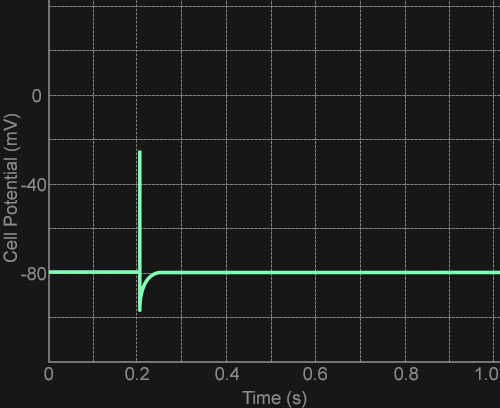
During an action potential, the sodium channels open quickly. Na+ ions rush into the neuron, making the voltage more positive. After a short delay, the sodium channels close and the potassium channels open. This brings the neuron back to its negative resting potential. The result is an upward voltage "spike" of a very short duration. This spike can be plotted on a graph of voltage against time, as shown here.
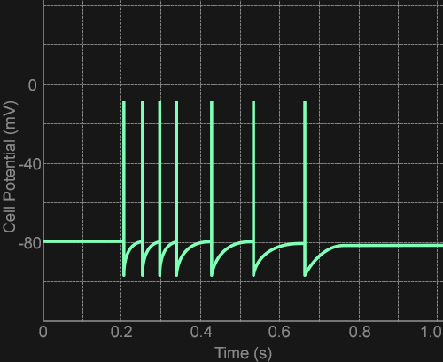
A typical sensory neuron transmits information about the intensity of a stimulus by generating different numbers of action potentials. For a weaker stimulus, a neuron may generate one action potential, as shown above. For a stronger stimulus, the neuron may generate multiple action potentials, as shown here.