Human Development
Human development involves the formation of different cells, tissues, and organs in an individual. are based on knowledge of the development of reproductive organs and other characteristics that are part of an individual’s .
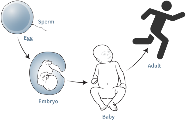
Human development starts with fertilization, the fusion of sperm and egg, and continues through birth and childhood.
Chromosomes
The development of reproductive organs in humans is set in motion by the actions of many genes, some located on the X and Y chromosomes. Most women have two X chromosomes, whereas most men have one X chromosome and one Y chromosome.
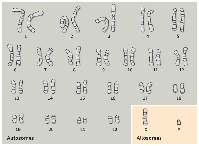
Humans have 22 pairs of and one pair of , for a total of 46 chromosomes. In most women, the two allosomes are both X. These women are referred to as 46,XX. In most men, the two allosomes are X and Y. These men are referred to as 46,XY.
X-Chromosome Inactivation
In every of individuals who are 46,XX, one of the two X chromosomes is and condenses into a compact structure called a Barr body. One type of sex verification test looks for the presence of a in somatic cells swabbed from inside an athlete’s cheek. The presence of a Barr body in each cell indicates that the individual has more than one X chromosome.

The Barr body can be seen as a darkly staining structure against the cell’s nuclear envelope. The other darkly stained body typically seen in the nucleus is a nucleolus, a nuclear subdomain where ribosomes are assembled.
Embryo
Until about 2 months after fertilization, the immature of human embryos are bipotential, meaning that they can develop into either or . The ultimate identity of the gonads results from the effects and interactions of many genes.
Urogenital Development (6 weeks)
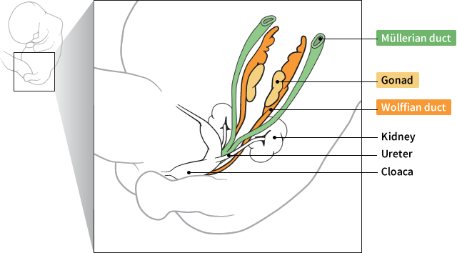
At 6 weeks, all human embryos have both Müllerian ducts (the precursors to the female reproductive system) and Wolffian ducts (the precursors to the male reproductive system).
Differentiation
One of the first genes that scientists identified as playing a role in sexual development is the sex determining region Y (SRY) gene. It affects a key control point for the differentiation of the immature gonads into testes. Absence of the SRY gene is one type of sex verification test for athletes to compete in women’s events.

Y Chromosome

The SRY gene is located on the Y chromosome.
Fetus
After about 9 weeks, the embryo becomes a fetus. Cells in the developing testes produce large amounts of that provide signals for the development of male internal and external reproductive anatomy. Cells in the developing ovaries produce large amounts of that promote the development of typical female anatomy.
Urogenital development (20 weeks)
Toggle between the female and the male fetus by selecting the bar.
Typical Female:

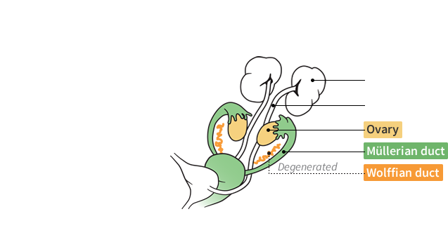
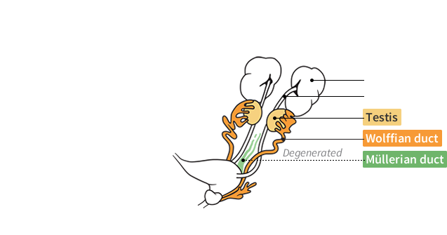
In the female fetus, the ovaries produce estrogens, which signal the Müllerian ducts to develop into female reproductive structures; the absence of testosterone signals the Wolffian ducts to degenerate. In the male fetus, the testes produce testosterone, which triggers the Wolffian ducts to develop into male reproductive structures; another hormone triggers the degeneration of the Müllerian ducts.
Birth
When a child is born, they are assigned a biological sex of female or male based primarily on the appearance of their or genitals. The physical examination of an athlete’s sex organs is one type of sex verification test.

Puberty
During puberty, the development of depends on the relative concentrations of and in the body and the body’s response to these hormones. Ovaries produce much more estrogens than testes do, and testes produce much more testosterone than ovaries do. Therefore, females typically produce more estrogens than males, whereas males typically produce more testosterone than females. Measuring blood testosterone levels is one type of test for athletes to compete in women’s events.
Blood Testosterone Levels in Elite Male and Female Athletes
Hide/unhide female and male levels by selecting “Female” and “Male”.


On average, male athletes have testosterone concentrations about 10 times those of female athletes, but there is a among individuals. (Source: Healy, M. L., et al. Clinical Endocrinology 81.2: 294-305, 2014.)
Phenotypes
Genetic variations can affect the typical development of reproductive anatomy and hormone levels in individuals, resulting in differences in sex development ( ). In most cases, individuals are still assigned a biological sex of female or male at birth.

Some of the more common DSDs are shown below.
Select the chromosome(s) for each variation to learn about the .
-
Typical biological female
-
CYP21A2 gene mutation
-
SRY gene mutation
-
AR gene mutation
-
SRY gene present
-
SRD5A2 gene mutation
-
AMH or AMHR2 gene mutations
-
Typical biological male
Select “Case Studies” to determine the biological sex of two fictitious athletes using the tests that have been employed throughout Olympic history.
Sex verification tests
Tests performed to verify an athlete’s biological sex have been referred to as sex-based tests, sex verification tests, gender tests, or gender verification tests (although these tests have to do with biological sex and not gender). They have only been conducted on female athletes in order to qualify them to participate in women’s events. More recently these tests have focused more on athletic performance than biological sex.
Biological sex
Biological sex is defined by an individual’s combination of chromosomes, hormone levels, internal and external reproductive anatomy, and sex characteristics. It is usually treated as a binary trait: female or male. Biological sex is different from gender, which is based on social and cultural ideas of what it means to be a woman or man. Gender identity refers to an individual’s concept of who they are. Gender identity does not always match biological sex.
Autosome
An autosome is any chromosome that is not an allosome (sex chromosome).
Allosomes
In humans, the allosomes are the X and Y chromosomes. The X chromosome spans about 155 million DNA base pairs and the Y chromosome about 59 million base pairs. Some textbooks refer to allosomes as "sex chromosomes," but the term is misleading because some genes on the X chromosome are involved in functions other than sex determination, whereas autosomes also contain genes involved in sex determination.
Somatic cell
Any cell of the body other than reproductive cells or gametes (e.g., egg and sperm).
Inactivated
One of the two X chromosomes is inactivated, which means that the genes on this chromosome are not expressed (e.g., do not produce RNA and proteins). X chromosome inactivation ensures that all individuals—female and male—have only one functional copy of the X chromosome in each cell. If inactivation did not occur, cells with two X chromosomes would express a potentially harmful double dose of certain genes.
Barr body
The inactive X chromosome forms a dense mass of DNA tightly wrapped around proteins. Which of the two X chromosomes in a cell is inactivated is random. The Barr body is named for the scientist who first discovered it: Canadian physician Murray Barr.
Gonads
The gonads are the primary reproductive organs: testes in males and ovaries in females. They produce the gametes (sperm or eggs) and sex hormones (androgens and estrogens).
Ovaries
The ovaries are a pair of female reproductive organs in which the eggs (ova) are produced. The ovaries are responsible for the production and release of sex hormones, primarily estrogens.
Testes
The testes are a pair of male reproductive organs. They are responsible for the production of sperm and sex hormones, primarily testosterone.
Testosterone
Testosterone is a type of steroid hormone called an androgen. It is produced by the testes in males and by the ovaries in females, and to a lesser extent by the adrenal glands in both sexes. Testosterone can also be taken illicitly by some athletes to increase athletic performance. This interactive focuses on testosterone naturally produced by the body.
Estrogens
Estrogens are a family of steroid hormones with a variety of functions. They are present in both males and females and are produced by the gonads. Estradiol is an estrogen that is important for the development of female reproductive organs and secondary sex characteristics. When most people talk about estrogen, they are referring to estradiol.
External sex characteristics
External sex characteristics include the opening of the vagina and the clitoris in females and the penis and scrotum in males. External sex characteristics are also called external reproductive anatomy, genitals, or genitalia. The appearance of external sex characteristics varies among individuals.
Secondary sex characteristics
Secondary sex characteristics include the physical characteristics caused by the surge in androgens and estrogens released during puberty. In males, they may include a deeper voice, more facial hair, increased muscle mass, and higher hemoglobin levels. In females, they may include enlarged breasts, widened hips, and an increase in body fat. There is variation among individuals in the appearance of these characteristics. The word "secondary" is used to distinguish these characteristics from those present at birth, such as genitalia, which are primary sex characteristics.
Range of values
Two large-scale studies of testosterone levels in athletes have been conducted, and they provide different results. The one shown here reported that 13.7% of female athletes have testosterone levels above the typical female range. The second study (Bermon, S. et al. J. Clin. Endocrinol. Metab. 99: 4328, 2014), which did not include any male athletes, reported that only 1.5% of female athletes have testosterone levels above the typical female range. Differences in how the measurements were taken and the athletes who were included may explain the differences in results.
DSD
Differences in sexual development are also referred to as intersex conditions or disorders of sex development. As many as 1 in 100 births show some differences from typical development.
Phenotype
The term phenotype refers to the observable characteristics or traits of an individual. Phenotype is the result of the interaction of the genetic makeup of an individual and environmental factors.
Typical biological female
In typical biological females, the SRY gene is absent. Phenotype: female internal and external reproductive anatomy and secondary sex characteristics, although there are variations among individuals (for example, variations in breast size or muscle mass); estrogens are typically much higher than testosterone, but there is variation in hormone levels and also in the body’s response to hormones.
CYP21A2 gene mutation
CYP21A2 mutations can alter the production of steroid hormones and cause enlargement of the adrenal glands, a condition known as congenital adrenal hyperplasia. Since the adrenal glands are a source of androgens, 46,XX individuals with this condition can produce excess testosterone and other hormones. Phenotype: ovaries present and functional; uterus present; enlarged clitoris; short vaginal canal; elevated testosterone levels (a condition referred to as hyperandrogenism); decreased fertility. Individuals are typically assigned to a female sex at birth.
SRY gene mutation
Mutations in the SRY gene may inactivate its function, and errors in chromosome segregation during meiosis may result in deletion of the SRY gene from the Y chromosome. Without a functioning SRY gene, the gonads do not differentiate into testes. Phenotype: uterus and fallopian tubes are present; ovaries are present but they do not produce eggs; typical female external sex characteristics. Individuals are typically assigned to a female sex at birth.
An error in chromosome segregation during meiosis results in only one complete X chromosome, with the other X chromosome being partially or completely missing. The condition is known as Turner syndrome. Phenotype: highly variable among affected individuals based on the extent and region of the missing X chromosome; female external reproductive anatomy; absent or reduced secondary sex characteristics; infertility; and ovaries may be absent or nonfunctional (e.g., do not produce eggs). Individuals are typically assigned to a female sex at birth.
AR gene mutation
To have an effect on cells, testosterone has to bind to a receptor called the androgen receptor (AR). Some mutations in the AR gene result in cells that don't respond to testosterone. The resulting condition is called androgen insensitivity syndrome. Phenotype: female or ambiguous external reproductive anatomy; male or ambiguous internal anatomy; breasts may develop. Individuals with complete androgen insensitivity syndrome are typically assigned to a female sex at birth, but there is a spectrum of phenotypes depending on the type of AR gene mutation involved.
An error in chromosome segregation during meiosis can cause an individual to have two X chromosomes and a Y chromosome. This condition is known as Klinefelter syndrome. Phenotype: male internal and external reproductive anatomy; low testosterone levels (hypoandrogenism); small testes; infertility; weak muscles; breasts may develop. Individuals are typically assigned to a male sex at birth.
SRY gene present
The SRY gene may be present on one of the X chromosomes as the result of crossing over between the X and Y chromosomes during meiosis. Phenotype: male external reproductive anatomy; small testes; low testosterone (hypoandrogenism); infertility. Individuals are typically assigned to a male sex at birth.
SRD5A2 gene mutation
Mutations in the SRD5A2 gene cause a deficiency in the enzyme 5-alpha reductase. Developing embryos do not produce enough of a hormone called dihydrotestosterone that is important for male sex development. Phenotype: male internal reproductive anatomy; female or ambiguous external reproductive anatomy at birth; surge in testosterone at puberty results in mostly male sex characteristics. Individuals may be assigned to either male or female sex at birth, but may reassign their sex to male after puberty.
AMH or AMHR2 gene mutations
In males, binding of the anti-Müllerian hormone (AMH) to its receptor, AMHR2, prevents the development of the Müllerian ducts into uterus and fallopian tubes. Mutations in the AMH and AMHR2 genes interfere with this process, resulting in the development of uterus and fallopian tubes. Phenotype: male internal and external reproductive anatomy, along with uterus and fallopian tubes; high testosterone; male secondary sex characteristics. Individuals are typically assigned to a male sex at birth.
Typical biological male
SRY gene is present. Phenotype: male internal and external reproductive anatomy and secondary sex characteristics, although there are variations among individuals (for example, variations in penis size, muscle mass, or facial hair); testosterone is typically much higher than estrogens, but there is variation in hormone levels and in the body’s response to hormones.