The stars of Bertozzi’s film were glycans: the sugar molecules that stud the outer surface of cells and are essential for important processes such as embryonic development, inflammation, and cancer progression.
To make the glycans visible under a microscope, researchers first fed cells chemically modified sugars that became incorporated into the glycans but did not affect their function. An imaging probe was then linked directly to the modified glycans. Bertozzi’s lab group at the University of California, Berkeley, is using the technique to monitor changes in the type, number, and distribution of glycans in cancer cells and in maturing cells. The researchers are also watching transparent zebrafish embryos as they develop.
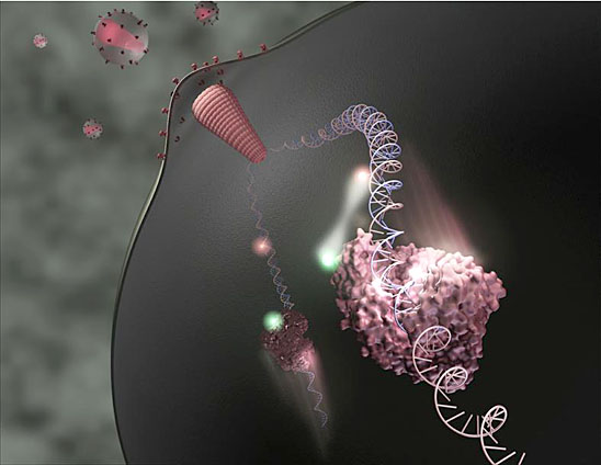
An acrobatic enzyme stole the show in the movie made by Zhuang at Harvard University. She tracked the precise movements of an HIV enzyme called reverse transcriptase, which spurs virus replication by converting viral genes into DNA that can integrate into the genome of an infected person. Zhuang used a technique called fluorescence resonance energy transfer, in which the transfer of energy between fluorescent dyes maps the proximity of two molecules, to illustrate how the enzyme zooms back and forth on the very DNA it is building.
She showed that when the enzyme falls off, which happens frequently, it grabs onto the middle of the DNA molecule and slides to the end to continue its work. If it finds that it is facing backward, it simply flips into the right orientation. Now, that’s entertainment.


