Virus Explorer


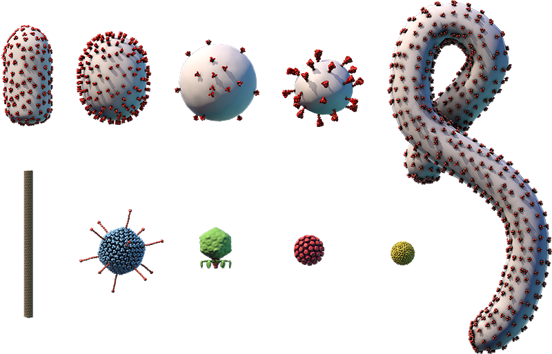
Click and drag or use the buttons below to move the model.
Use the tab to focus one of the buttons above, and then space to start the rotation.
The proteins embedded in the viral envelope are shown.
Components on the virus are selectively represented in the 3D model for educational purposes.

A. Envelope protein; B. Membrane protein; C. Lipid envelope; D. RNA genome; E. Capsid protein

This diagram shows how Zika virus replicates, or makes copies of itself. Glycoproteins on the surface of Zika virus bind to specific receptors on the host cell’s surface (A). This triggers a process called endocytosis (B), which brings the virus into the cell in an enclosed structure (vesicle) called an endosome. Acidic substances build up inside the endosome, lowering the pH and triggering the endosome’s membrane to fuse with the virus’s envelope. This releases the virus’s (+)RNA genome into the cell’s cytosol (C).
The cell’s ribosomes translate the virus’s genome into one large protein (D) that is cleaved, or cut into smaller proteins, including a viral polymerase (E). The viral polymerase transcribes the virus’s (+)RNA genome into a complementary (–)RNA template (F). This template is used to make copies of the virus’s whole (+)RNA genome (G).
Viral genomes and proteins assemble into new viruses on the endoplasmic reticulum (ER). The new viruses travel through the ER-Golgi network through a process called budding. This process surrounds the virus in a piece of the cell membrane containing viral proteins, which becomes the virus’s envelope. The viral proteins are further modified through protein processing (H). Once a new virus becomes fully formed through a series of changes called virus maturation (I), it is released from the cell in a process called exocytosis (J).
Zika Virus
- Part of the Flaviviridae family, which also includes yellow fever virus, West Nile virus, and dengue virus
- ~50-nm enveloped particles with surface proteins that form an icosahedral structure surrounding the envelope
- Linear (+)ssRNA genome of ~10,800 bp
- Infects humans and other primates
- Common vectors are mosquitoes of the genus Aedes, which carry and transmit the virus between hosts
- No vaccine currently available
Zika virus is one of several mosquito-borne flaviviruses. The virus is spread primarily through the bite of Aedes aegypti and related species of mosquitoes.
The virus was of limited concern until 2015, when scientists in South America discovered an association between Zika virus infection in pregnant women and an increase in the number of babies born with smaller-than-normal head sizes, a condition known as microcephaly. Scientists have since agreed that Zika virus infection in pregnant women may lead to microcephaly and other neurological defects in their children.
Historical Timeline of Zika Virus Spread

Zika virus’s date of first appearance
- 1947–1952
- 1954
- 1969–1983
- 2012–2014
- Feb 2015
- Oct 2015
- Nov 2015
- Dec 2015
- Jan 2016
- May 2016