Virus Explorer


Click and drag or use the buttons below to move the model.
Use the tab to focus one of the buttons above, and then space to start the rotation.
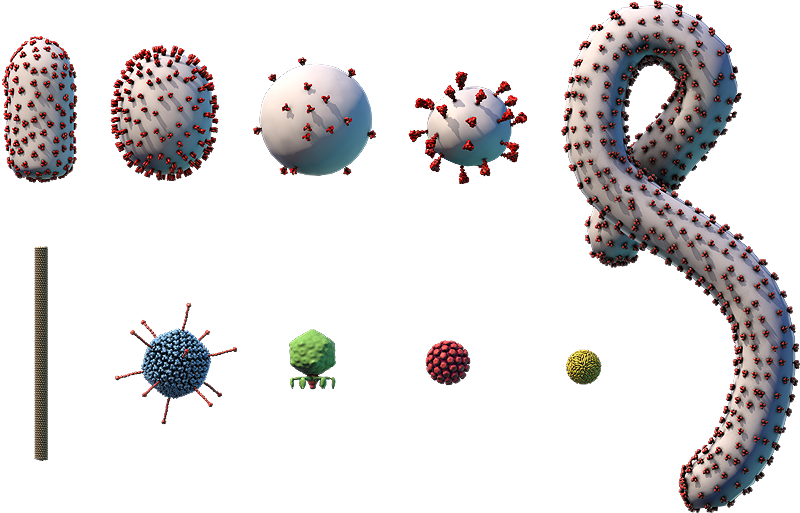
The viral envelope (gray) and proteins embedded in the envelope (red) are shown.
Components on the virus are selectively represented in the 3D model for educational purposes.

A. Glycoprotein; B. Lipid envelope; C. Matrix protein; D. RNA genome; E. Nucleocapsid proteins; F. Polymerase

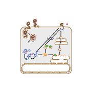
This diagram shows how Ebola virus replicates, or makes copies of itself. Glycoproteins on Ebola virus’s surface bind to specific receptors on the host cell’s surface (A). This triggers a process called endocytosis (B), which brings the virus into the cell in an enclosed structure (vesicle) called an endosome. Acidic substances build up inside the endosome, lowering the pH and triggering the endosome’s membrane to fuse with the virus’s envelope. This releases the virus’s (–)RNA genome and viral polymerase proteins into the cell’s cytosol (C).
The viral polymerase transcribes the virus’s (–)RNA genome into a complementary (+)RNA template (D). This template is used to make copies of the virus’s whole (–)RNA genome (E).
The viral (–)RNA is also transcribed into mRNAs (F), which are translated into proteins by ribosomes in the cytosol and on the endoplasmic reticulum (ER) (G). Ebola virus glycoproteins go through protein processing in the ER-Golgi network (H), then are transported to the cell membrane.
Viral genomes and proteins are assembled into new viruses (I), which leave the cell through a process called budding (J). This process surrounds the virus in a piece of the cell membrane containing viral proteins, which becomes the virus’s envelope.
Ebola Virus
- Part of the Filoviridae family, which also includes Marburg virus
- ~80-nm × 800-nm enveloped particles with a helical capsid
- Linear (–)ssRNA genome of ~19,000 bp
- Infects bats, humans, and other primates
- First vaccine against one species of Ebola virus was approved for adults in 2019
Ebola virus (and its relative Marburg virus) is perhaps best known for its gruesome symptoms and high death rate in humans. This virus is zoonotic, which means it can be transmitted to humans from infected animals, such as bats.
Outbreaks of Ebola virus infections occurred in Africa as early as 1976. But until 2014, they were short-lived and isolated to relatively small groups of people. In 2014, an Ebola outbreak in West Africa spread primarily across Sierra Leone, Guinea, and Liberia, killing more than 11,000 people over the course of two years. During this time, the virus also spread in several other countries, but was limited and quickly controlled.
Historical Timeline of Ebola Virus Outbreaks
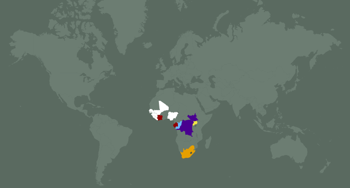
Ebola virus’s year of first appearance
- 1976
- 1994
- 1996
- 2000
- 2001
- 2014