Anatomy of p53
p53 contains several domains (protein parts).
The transactivation domain (red) allows p53 to activate other genes after binding to their regulatory regions. The domain recruits RNA polymerase and other enzymes that transcribe RNA.
The DNA binding domain (green) is responsible for p53’s ability to bind to the regulatory sequences of genes. Most mutations in the p53 protein found in cancers are in this domain.
The complexing domain (yellow) is responsible for bringing four individual p53 molecules together.
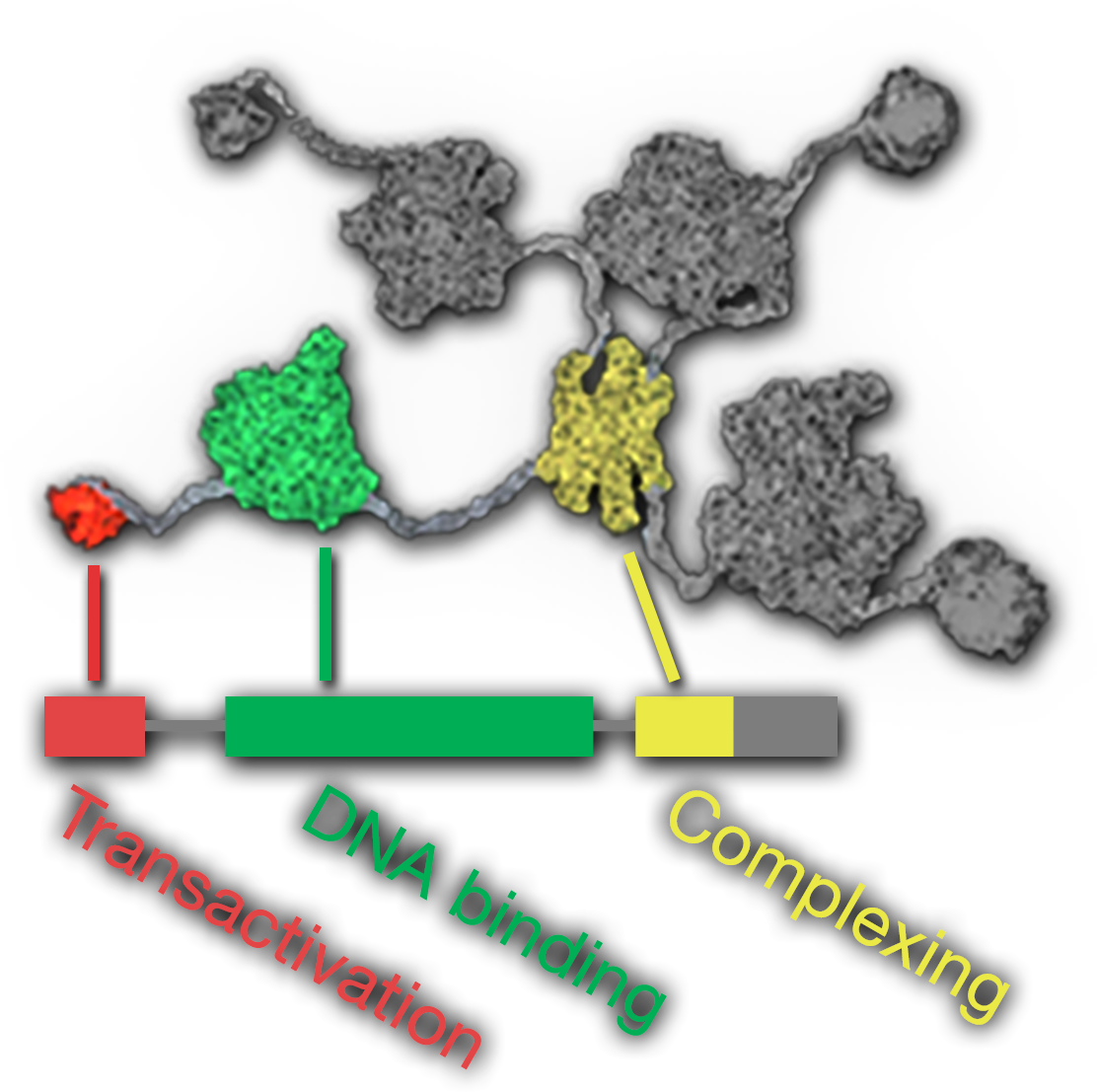
A depiction of a molecule as a central yellow blob with four gray tails extending from it. About midway along each tail is a large blob. Three of these blobs are gray and one is green. At the end of each tail is a small blob. Three of these blobs are gray and one is red. Below this structure is a series of colored rectangles of different lengths labeling various parts of the image. The red blob is labeled Transactivation, the green blob is labeled DNA binding, and the yellow blob is labeled Complexing.