Virus Explorer
Click and drag or use the buttons below to move the model.
Use the tab to focus one of the buttons above, and then space to start the rotation.
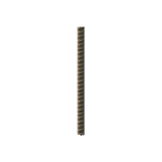
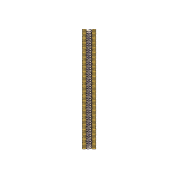
The proteins that make up the helical capsid are shown.
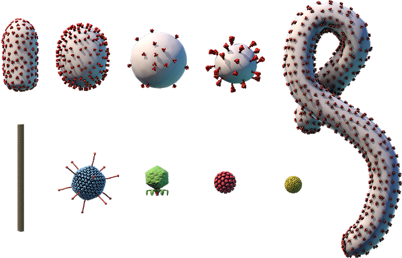
Components on the virus are selectively represented in the 3D model for educational purposes.

A. Coat protein; B. RNA genome

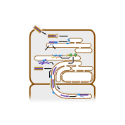
Replication cycle legend: This diagram shows how TMV replicates, or makes copies of itself. TMV enters a plant cell through a wound in the cell wall (A). The virus’s outer coat protein disassembles (B), releasing the (+)RNA genome into the cell’s cytosol.
Ribosomes on the endoplasmic reticulum (ER) translate viral replication proteins from the virus’s (+)RNA genome (C). The replication proteins transcribe the virus’s genome into a complementary (–)RNA template (D). This template is used to make copies of the virus’s whole (+)RNA genome (E) and is also transcribed into mRNAs (F).
The mRNAs are translated into viral proteins by ribosomes on the ER (G). These proteins include coat proteins, which are used to build new viruses that may exit and enter new cells through wounds in the cell wall (H), and movement proteins, which help the viruses infect adjacent plant cells. Plant cells are connected by a large ER network that passes through tiny openings in their cell walls, called plasmodesmata. The movement proteins interact with host cell proteins to transport viral genomes and replication proteins along this ER network, through the plasmodesmata, and into adjacent plant cells (I), where the virus continues to replicate (J).
Tobacco mosaic virus (TMV)
- Part of the Virgaviridae family
- ~18-nm × 300-nm naked helical capsid
- Linear (+)ssRNA genome of ~6,500 bp
- Infects plants such as tobacco and tomato plants
- No vaccine, but scientists have produced genetically engineered plants that are resistant to infection
Tobacco mosaic virus (TMV) was the first virus to be identified and described. Its name comes from the mosaic-like pattern it causes on the leaves of infected plants.
Studies of TMV — including the purification of the virus in the late 1800s, followed by the characterization of the virus in the early 1900s — laid the foundation for the field of virology. Today, many plants have been genetically modified to resist infection by TMV.